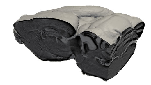
A virtual cut through the tomographic data set, reconstructed from phase-contrast measurements. The technique allows one to look inside the brain and see the plaques, without actually cutting the brain up into slices. (Figure: Paul Scherrer Institut/B. Pinzer)
Swiss researchers have succeeded in generating detailed three-dimensional images of the spatial distribution of amyloid plaques in the brains of mice afflicted with Alzheimer’s disease. These plaques are accumulations of small pieces of protein in the brain and are a typical characteristic of Alzheimer’s.
The new technique used in the investigations provides an extremely precise research tool for a better understanding of the disease. In the future, scientists hope that it will also provide the basis for a new and reliable diagnosis method. The results were achieved within a joint project of two teams of researchers – one from the Paul Scherrer Institute (PSI) and ETH Zurich, the other from the École Polytechnique Fédérale de Lausanne (EPFL). They have been published in the journal Neuroimage.
Alzheimer’s disease is responsible for about 60% to 80% of all cases of dementia. This disease affects people differently, but the most common initial symptom is the difficulty in remembering new information, because the disease first affects brain regions involved in the formation of new memories. Alzheimer’s dementia is characterized by typical brain lesions that spread to other brain regions as the disease progresses. One of these lesions, the so-called amyloid plaque, is composed of the accumulation of extracellular protein aggregates. These lesions appear early in the course of the disease and there is a high interest in detecting them in patients to diagnose or evaluate the progression of the disease. Recently, medical imaging methods have been developed and validated for this purpose. These allow regional amount of amyloid deposits to be measured, but individual plaques cannot be quantified. The latest results obtained by researchers from the Paul Scherrer Institute (PSI), ETH Zurich and the École Polytechnique Fédérale de Lausanne (EPFL) show that imaging single plaques is feasible under certain conditions. “This achievement could help to advance the development and evaluation of new imaging diagnostic markers for ultimately improving the diagnosis of Alzheimer’s disease,” explains Matthias Cacquevel, one of the authors at EPFL.
